HEEL PAIN

DO YOU WANT TO WATCH A SHORT VIDEO WHICH DR. TJ AHN EXPLAINS WHAT HEEL PAIN IS AND HOW TO FIX IT?

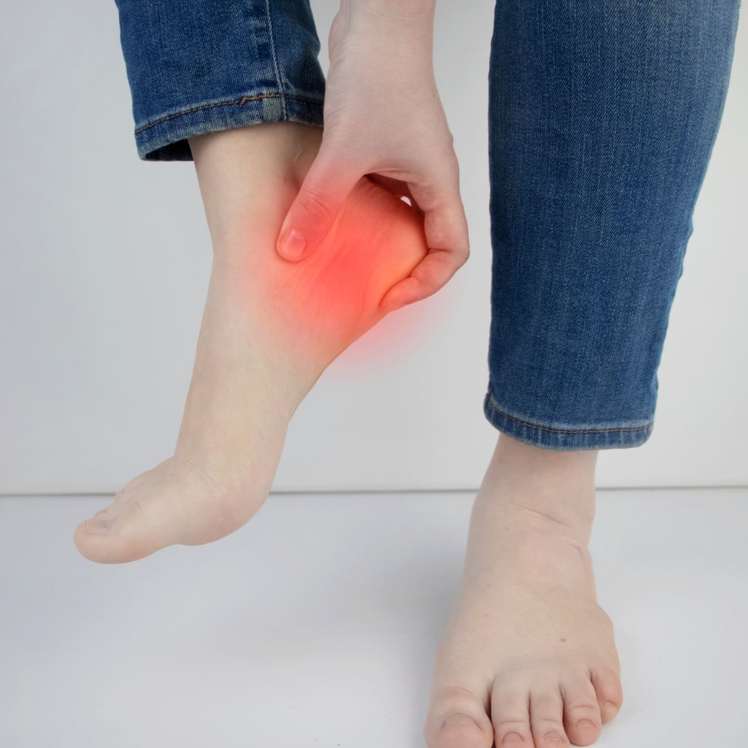
Heel pain is most often caused by plantar fasciitis, a condition that is sometimes also called heel spur syndrome when a spur is present.  Plantar fasciitis is an inflammation of the thick band of fibrous connective tissue (fascia) running along the bottom (plantar surface) of the foot, from the heel to the ball of the foot.

ANATOMY

Hammertoes can be flexible or rigid in nature. If they are rigid, it is not possible to straighten the toe out by manipulating it. They tend to slowly get worse with time and often flexible deformities become rigid.
Frequently hammertoes develop corns or calluses (a buildup of skin) on the top, side, or end of the toe, or even between two toes. These may be soft or hard, depending upon their location. Corns and calluses can be painful and make it difficult to find a comfortable shoe, but even without corns and calluses, hammertoes can cause pain because the joint itself may become dislocated.
CAUSES

Biomechanics: Hammertoes develop mainly from abnormal foot structure and mechanics, both of which are inherited features. There are some inherited foot defects such as flat feet and high arches that will put excessive strain on the muscles and tendons making them more susceptible to hammertoe deformity over time.

Footwear: Certain types of footwear such as high heeled or tight, ill-fitting shoes can contribute to the development of hammertoes. That is why hammertoes are more common in women than men. Improper footwear can exacerbate the problem caused by the original genetic structure. Tight shoes force the toes to stay in a bent position for too long, restricting the muscles ability to flex, and tendons to shorten over time, causing tightening and making it more difficult to straighten the toe.

Injury: Injuries to the foot can also be a factor in developing a hammertoe. A broken bone in the toe or foot may heal improperly or cause damage to the muscle, tendon or joint, causing a hammertoe.

Physical Conditions: People suffering from diabetes, arthritis, gout, poor circulation, brain, spinal cord or
nerve injuries such as stroke, cerebral palsy and degenerative disc disease are at an increased risk of developing a hammertoe.

Symptoms

Pain: The pain could be most prominent when footwear is being worn, but it can also continue once the shoe is removed. The pain may be evident only when the toe is moved as in walking, but it can also become constant. Pain in the ball of the foot at the base of the affected toe is also common with hammertoes.

Corns and Calluses: Early signs of hammertoe are often corns or calluses that form on the affected toe. When the toe starts to bend into the hammer shape, a corn or callus can develop on the top of the toe or beneath the toe near its base, where it rubs against the shoe or even between the toes. A thickening of the skin due to undue pressure or constant rubbing causes both corns and calluses.

Restricted Motion: The affected toe or toes may become stiff and hard to move over time.

Redness and swelling: This can occur at the joint contracture. In severe cases, ulceration and infection can develop. The patient usually seeks treatment for painful and deformed toes, associated with calluses on the affected toe joints.

Diagnosis

Physical exam: Clinical observation of the hammertoe is typical to make the diagnosis. Your doctor will ask you questions about the symptoms you are experiencing. The structure and biomechanics of the patient’s entire foot is examined.

X-Rays: X-rays are necessary to get a better understanding of the extent of the bone deformity and contracture.

Treatment

Non-Surgical Treatment:

The first method of treating hammertoes begins with accommodating the deformity, and is indicated in mild deformities and functional abnormalities. The goal is to reduce friction and relieve pressure on the painful area.

Padding: Gel pads recommended by your doctor can help prevent irritation to corns and calluses that have developed from the hammertoe.

New Shoes: Avoiding shoes with pointed toes or high heels and shoes that are too short in the toe box will prevent the hammertoes from being forced against the front or top of your shoe. Comfortable shoes with a wide and roomy toe box and short to no high heels will offer more support and comfort to the toes.

Orthotic devices: A functional orthotic device may be prescribed by your doctor to be worn in your shoes to help control the muscle/tendon imbalance, bringing pain relief.

Injection therapy: Corticosteroid injections are sometimes used to ease the inflammation associated with hammertoes.

NSAIDS: 0ver the counter oral non-steroidal anti-inflammatory drugs (NSAIDs), such as ibuprofen may be recommended to reduce pain and inflammation.

Splinting/strapping: Your doctor may apply splints or small straps for temporary relief. This is more common with flexible types of hammertoes.

Stretching: Flexing and stretching the muscles and gently massaging may alleviate some of the pain and help relax the muscles and tendons.

Soaks and Moisturizers:

Soak the foot in warm water mixed with Epsom salts and apply an appropriate moisturizing cream to help keep the skin soft.

Conservative treatments for hammertoes are often limited because they cannot correct the bone deformities involved. There is no way to stop the progression or reverse the deformity without literally moving the bones back into the correct position and realigning the joint. This can only be accomplished with surgery. If conservative treatment fails or the hammertoes progress to the point where conservative treatment is no longer a viable option, surgical intervention may be needed to correct the deformity.


Surgical Treatment

Hammertoe Surgery

Both techniques involve performing arthroplasties and correction of the deformity involves working on the bones and soft tissues. In the traditional approach, range of motion is limited or absent due to the fusion of the joints. In the minimally invasive technique, one of the goals is to preserve the joints and as a consequence, range of motion is usually maintained. Because the minimally invasive techniques are less traumatic, and the recovery time shorter, most patients prefer this

method of correction. Each patient is unique, however, and the doctor will discuss which technique he or she feels best suits the patient for optimal outcome.

This strong and tight tissue contributes to maintaining the arch of the foot. It is also one of the major transmitters of weight across the foot as you walk or run. Therefore, the stress placed on this tissue is tremendous. This condition is one of the most common complaints relating to the heel. The pain is usually felt on the underside of the heel and is often most intense with the first steps of the day, or first step after a long period of being off your feet. 

The condition occurs when the plantar fascia is strained over time beyond its normal extension, causing the soft tissue fibers of the fascia to tear or stretch at points along its length; this leads to inflammation, pain, and possibly the growth of a bone spur where it attaches to the heel bone.

A heel spur is a bony growth that occurs at the attachment of the plantar fascia to the heel bone (calcaneus). A heel spur can be present on a foot with no symptoms at all and a painful heel does not always have a heel spur present.

CAUSES

Biomechanical factors: The most common cause of plantar fasciitis relates to faulty structure of the foot. Over-pronation (flat feet) is the leading cause of plantar fasciitis. Over-pronation occurs in the walking process, when a person’s arch collapses upon weight bearing, causing the plantar fascia to be stretched away from the heel bone. High arches and tight tendons along the back of the heel (Achilles tendon) are also structural foot abnormalities that can lead to plantar fasciitis.

Aging: The natural process of aging, which may cause tissue in the heels to weaken over time and/or promote wear and tear is sometimes a factor in the development of plantar fasciitis.

Obesity or sudden weight gain: Excess pounds put extra stress on your plantar fascia

Footwear: Non-supportive footwear puts abnormal strain on the plantar fascia and can also lead to plantar fasciitis. Improper footwear can exacerbate the problem caused by the original genetic structure.

SYMPTOMS

The most common complaint is pain and stiffness in the bottom of the heel. Pain is often at its worst first thing in the morning. Often the pain disappears after the first steps and can reappear after standing or walking for long periods. The heel pain may be dull or sharp and the bottom of the foot may also ache or burn. Mild swelling is possible and there may be difficulty bending the toes towards the shin (called “dorsiflexion” of the foot).

DIAGNOSIS

Physical exam: Clinical observation is typical to make the diagnosis. Your doctor will ask you questions about the symptoms you are experiencing. The structure and biomechanics of the patient’s entire foot is examined.

X-rays: Standard weight-bearing radiographs in the lateral and anteroposterior projection demonstrate the biomechanical character of the hindfoot and forefoot, and may show other osseous abnormalities such as fractures or rheumatoid arthritis in the calcaneus. Radiographs serve as an aid to confirm the clinician’s diagnosis.

TREATMENT

NON-SURGICAL TREATMENT

Stretching exercise: Exercises that stretch out the calf muscles help ease pain and assist with recovery. Try gentle calf stretches for 20 to 30 seconds on each leg. This is best done barefoot, leaning forward towards a wall with one foot forward and one foot back. Your doctor can give you recommendations for appropriate stretching exercises or in some cases may refer you to a physical therapist.

Appropriate shoes: Wearing supportive shoes that have good arch support and a slightly raised heel reduce stress on the plantar fascia. Wear shoes that fit well and consider purchasing shoes with shock-absorbent soles, rigid shanks, and supportive heel counters. Avoid going barefoot because walking without shoes puts undue strain and stress on your plantar fascia.

Ice: Putting an ice pack on your heel for 15 minutes several times a day helps reduce inflammation. Place a thin towel between the ice and your heel; do not apply ice directly to the skin.

Activities: Cut down on extended physical activities to give your heel a rest. Avoid running on hard surfaces and other high impact sports.

Medications: Oral non-steroidal anti-inflammatory drugs (NSAIDs), such as ibuprofen, may be recommended to reduce pain and inflammation.

Padding and strapping: Placing heel cushions in the shoe softens the impact of walking. Strapping helps support the foot and reduces strain on the fascia.

Orthotic devices: Custom orthotic devices that fit into your shoe may be prescribed for correcting biomechanical imbalance, controlling excessive pronation, and supporting the ligaments and tendons attached to the heel bone. Orthotics can effectively treat many cases of heel and arch pain without the need for surgery.

Shockwave Therapy: Radial Pulse Shockwave Therapy is an FDA cleared technology proven to increase the rate of healing for soft tissue injuries and for the relief of painful conditions of the foot and ankle. It is the most advanced non-invasive and highly effective treatment method that enhances blood circulation, accelerates the healing process causing damaged tissue to gradually regenerate and heal. Click here to learn more about this treatment option.

Stem Cell Recruitment Injection therapy: In some cases, corticosteroid injections are used to help reduce the inflammation and relieve pain. However, stem cell recruitment therapy with 100% pure amniotic fluid injection is a great choice since these safe, drug-free injections contain growth factors and other naturally occurring nutrients which encourage cellular and soft tissue regeneration and reconstruction. Click here to learn more about this treatment.

Removable walking cast: A removable walking cast may be used to keep your foot immobile for a few weeks to allow it to rest and heal.

Night splint: Wearing a night splint allows you to maintain an extended stretch of the plantar fascia while sleeping. This may help reduce the morning pain experienced by some patients.

Although most patients with plantar fasciitis respond to non-surgical treatment, a small percentage of patients may require surgery. If, after several months of non-surgical treatment, you continue to have heel pain, surgery will be considered.

SURGICAL TREATMENT

Plantar Fasciitis and Heel Spur Surgery

Both the traditional and minimally invasive surgical correction techniques are performed at the United Foot & Ankle Surgeons. Both techniques involve relieving the tension on the plantar fascia (plantar fasciotomy or cutting of the fascia) and relieving the pain of heel spur syndrome which often involves removing the spur also.

Because the minimally invasive techniques are less traumatic, and the recovery time shorter, most patients prefer this method of correction. Each patient is unique, however, and the doctor will discuss which technique he or she feels best suits the patient for optimal outcome.

Traditional Plantar Fasciitis and Heel Spur Surgery

Open or traditional surgical correction techniques involve a large incision, several centimeters in length, which is made on the inside of the heel. This allows the surgeon access to the spur and the connection of the plantar fascia to the heel bone. In removing the spur from the heel, the plantar fascia is partially cut and released from its connection to the heel bone. This releases the tension in the plantar fascia, thus relieving the patient’s pain. The incision is closed with stitches, and you are placed in a surgical bandage to protect the foot while it heals. You may be placed in a splint for certain open technique corrections. The large incision into the heel precludes, in some cases, the patient from bearing any weight on the foot for 2-4 weeks.

Minimally Invasive Plantar Fasciitis and Heel Spur Surgery

The technique used in minimally invasive or minimal incision percutaneous surgery involves a much smaller incision of the heel for release of the fascia and removal of the spur if present. It involves making a small incision less than 1 cm to release the plantar fascia, which may be combined with heel spur removal. Suture is usually not necessary. For this technique, surgeons use instruments which are 

very fine and rotate at high speed to make tiny, precise cuts. Surgery is performed under Fluoroscopic viewing. There is less trauma to the tissues and surgical times are lessened with this technique, reducing pain and recovery time. Postoperative patients ambulate immediately and are often placed in a surgical shoe or boot to aid ambulation. The most commonly performed MIS procedure for correction of heel spur syndrome is the Isham modified Schwartz procedure.

Please call us at 773-989-2500 or click below to schedule an appointment online!